
Exploring the Anatomy of a Horse: Printable Parts for Education and Fun


Understanding the External Anatomy of a Horse
Horses are magnificent creatures that have been a part of human history for thousands of years. With their impressive strength, agility, and beauty, it's no wonder why many people are fascinated by these animals. For horse enthusiasts, students, and educators, understanding the anatomy of a horse is essential for appreciating their biology and behavior. One way to learn about the different parts of a horse is through printable diagrams and resources.
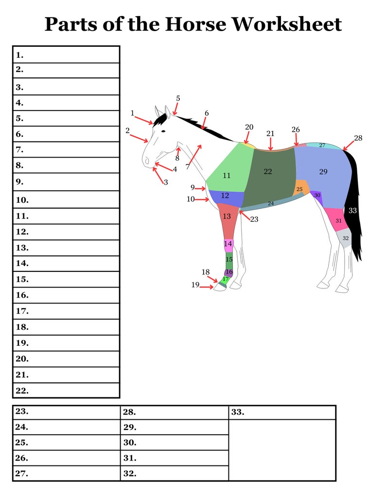
The external anatomy of a horse includes various parts such as the head, neck, body, legs, and tail. Each of these parts plays a crucial role in the horse's overall health and well-being. For example, the head contains the brain, eyes, ears, and mouth, which are all vital for the horse's senses and communication. The legs, on the other hand, provide support and enable the horse to move around. Printable diagrams of a horse's external anatomy can help individuals learn and identify these different parts.
Internal Organs and Systems of a Horse
In addition to the external anatomy, it's also important to understand the internal organs and systems of a horse. The internal organs include the heart, lungs, liver, and kidneys, which all work together to keep the horse alive and healthy. The digestive system, which includes the mouth, esophagus, stomach, and intestines, is responsible for breaking down food and absorbing nutrients. Internal Organs and Systems of a Horse
Printable parts of a horse can be a valuable resource for educators, students, and horse enthusiasts. These diagrams can be used to create educational materials, such as worksheets, posters, and study guides. They can also be used to create fun and interactive activities, such as puzzles, quizzes, and games. By learning about the different parts of a horse, individuals can gain a deeper appreciation for these amazing animals and develop a stronger understanding of their biology and behavior.